What you should know before exploring the rest of this site
Terminology
- Allele: A form of a gene caused by different bases at certain points within a gene. One allele is received from each parent, and there is often more than two alleles per gene.
- Chromosomes: Thread-like structures located inside the nucleus of animal and plant cells.
- Deoxyribonucleic Acid (DNA): Genetic material coding for the formation of an organism as instructed by the order of its nucleotides.
- DNA probe: A radioactively labeled single strand of nucleic acid molecule used to tag a specific sequence in a DNA sample.
- DNA Sequencing: The process of finding out the order of nucleotide base pairs in a length of DNA
- Double Helix: A twisted ladder shape consisting of two strands that are anti-parallel to each other.
- Gene: Heritable units parents pass on to offspring
- Human Genome: The comprehensive genetic information of a human.
- Locus (plural Loci): the location of a gene on a chromosome
- Nucleotides: A composition of a nitrogenous compound, a phosphate, and deoxyribose. There are four types of nucleotides in DNA
- Restriction Enzyme: An enzyme that is able to ‘cut’ or digest this DNA into smaller pieces.
- X-Ray Crystallography: A technique used to determine the 3D structure of a molecule.
 The unique 'X' structure of chromosomes keeps DNA tightly packaged. Without such wrapping, DNA molecules would be too long to fit inside cells. Supposedly, if all of the DNA molecules in a single human cell were uncoiled and placed end-to-end, they would stretch 6 feet. A human somatic cell has 46 chromosomes in total. 44 of these chromosomes are autosomes (chromosomes not involved in sex determination). The remaining two chromosomes are sex chromosomes. Females have two X chromosomes, and males have one X and one Y chromosome.
 DNA Model
In 1953, Watson and Crick proposed the double helix structure of DNA. They amassed data from other scientists in order to build the DNA model. 2 crucial information sources were from Rosalind Franklin's X-Ray crystallography of DNA, which indicated that DNA was in the shape of a double helix, and Erwin Chargaff's knowledge of nucleotides and their pairing patterns, now known as Chargaff's Rules.
DNA is a polymer of repeating units of nucleotides, which consist of a 5 carbon sugar (deoxyribose), a phosphate, and a nitrogen base. The 4 nitrogenous bases are Adenine (A), Thymine (T), Guanine (G), and Cytosine (C). A bonds to T by a double hydrogen bond. C bonds to G by a triple hydrogen bond.

Restriction Enzymes
A restriction enzyme is also called an endonuclease because it cuts within ("endo-") the DNA instead of at the ends ("exo-"). Every restriction enzyme digests DNA at a designated site defined by a sequence of bases in the DNA called a recognition site. “Sticky ends” are single stranded ends of a fragment which can pair with another sticky end that has a complimentary sequence.
Gel Electrophoresis
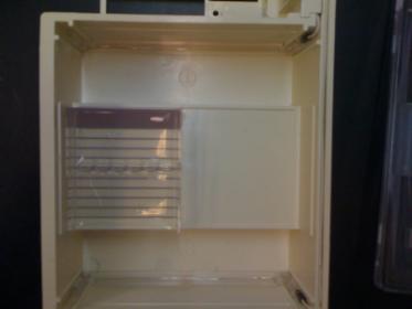
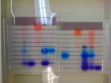
Gel electrophoresis is a technique used to determine the size of different strands of DNA. It sorts the DNA strands through electricity. Since DNA is negatively charged due to the phosphate that is part of the backbone, the wells in the agarose gel are situated near the negative end of the box. While the electricity is running, the negatively charged DNA strands are attracted to the positive end of the gel. The agarose gel the DNA must move through is a matrix, which means larger strands get caught up in the matrix and move through the gel much more slowly than the smaller fragments. The picture on left below shows the gel box and gel. In the case of this picture, dyes were used which had positive and negative charges, so the wells were located in the center of the gel. The image on the bottom right shows how the dyes traveled while the electricity was running. The top of the picture is the negative end, so the red dye was positively charged. The purple dye traveled farther than the blue dye, which indicates that the purple dye was composed of smaller molecules than the blue dye.
 |  |